55:247 Image Analysis and Understanding

Project Listing - Spring 2014


IP = Image processing part

AI = Knowledge-based part


Name: Jason Agne

Image Data

Fundus and OCT image pairs


Goals

Predict ONH volume using Fundus and OCT features in combination.

(what exactly do you mean by volume? cup? rim? both?)


Methods I, IP

Extract vessels, detect ONH rim, and compute image features of OCT/ONH image from both fundus and OCT. Apply a regression algorithm. Start from there and perhaps extract more elaborate features. Apply PCA to eliminate unneeded ones.

Methods II, AI

OCT segmentation used to compute 'true' volume.

Explore whether these feaures can predict volume - not using any features directly corresponding to volume.

Validation

Compare predicted volume with ONH volume calculated from segmentation

Use 20 image pairs of OCT/fundus, make sure training and testing are completely separated.

Name: HanQin Cai

Image Data

Street view pictures including traffic signs with natural background.

Goals

Recognizing the traffic signs from the nature street view pictures.

Methods I, IP

Considering the common designs of the traffic signs, using color-based method and shape-based method to detect traffic signs from the picture.

Methods II, AI

Reading the texts and symbols from the traffic signs, then comparing with a traffic signs database to classify the traffic signs. If necessary, also figure out the additional information from the signs (e.g. the number of the speed limit).

Validation

Processing at least 60 pictures with different combinations of signs, different weather (e.g. sunny, cloudy, rainy, night), and different backgrounds (e.g. trees, buildings, and highway). Then comparing the accurate results, the accurate results can be easily observed by human, no professional knowledge required.

Name: Jiawen Chen

Image Data

3D OCT images from subjects with Age-related Macular Degeneration

Goals

1) Identify rough locations of SEADs = blood filled cysts in the retina

2) Determine accurate surfaces of these SEADs

Methods I, IP

Segmentation using mean-shift approaches, or Viola-Jones approaches (integral image features and boosting), or random forest approaches

Methods II, AI

Utilize existring 11-layer segmentation of retinal layers to help you identify what is and what is not a SEAD.

Validation

Use existing independent standard with SEADs traced in 3D, compare in at least 90 images from OPTIMA project or Iowa-AMD project. For Goal 1, validate to determine how many SEADs were correctly identified, missed, and falsely detected where no SEADs are present. For Goal 2, use Dice coeeficients and perhaps surface positioning errors to determine accuracy.

Name: Ali Ghayoor

Image Data

MRI-T1 scans of human brain acquired from 8 different sites using 4 different scanners including SIEMENS TrioTim and Verio, Philips Intera Achieva, and a GE Signa HDxt.

Goals

Develop a method for automated design of optimal or at least good metrics that can be used for image registration via non-linear optimization.

Fast diffeomorphic image registration using non-linear optimizers

Methods I, IP

First, a common non-linear optimizer like L-BFGS-B or Ipopt (interior point optimizer) will be implemented in ITKv4; then, a new optimizer will be designed based on the above methods to handle displacement field transforms optimization.
Finally the designed filter will be applied for SyN registration in ANTs (Advanced Normalization Tools) that currently uses conjugate gradient descent linear optimizer for high diffeomorphic registrations.


Methods II, AI

Although the linear optimizers work well for low-dimensional registrations (e.g. Affine, rigid), non-linear optimizers are much faster for dens registration and generally when the parameter space is big (like BSpline registration).

The family of quasi-newton methods is popular, but these iterative nonlinear optimizers cannot handle the transforms with local support like displacement field transforms, so they cannot be used in high diffeomorphic registration (like SyN). It happens because:

1- these optimizers keep a history of previous updates to estimate the Hessian matrix from the successive gradient vectors.
2- in diffeomorphic registration, the “regularization” process changes the optimizer outputs, so it causes conflicts with the values that are saved in optimizer internally for the estimation of Hessian matrix.

Although the above issue can be avoided if the optimizer is fed with Hessian matrix before each iteration, computing the Hessian matrix is almost impractical for dense transforms like displacement field transforms.
To come up with this problem, the Hessian matrix estimation can be transferred to one level up outside of the optimizer. Having the regularization outputs the Hessian matrix can be updated using a method that is used in Quasi-Newton algorithms.
By doing the estimation outside of the optimizer, it does not need to keep track of the previous values internally, so the conflict issues will be fixed.

After the implementation of the above method, we can compare the accuracy and the speed of the registration process to the case when linear methods are used for optimization.

Validation

Run displacement field image registration using conjugate gradient optimizer and the designed non-linear optimizer on 30 multi-site, multi-scanner MRI-T1 images of the human brain, and compare the accuracy, robustness and the running time of the results.


Name: Melanie Kneisel

Image Data

BMP, 256 gray-levels, uncompressed fingerprint images will be used from the Fingerprint Verification Competition (FVC) databases. Each year organizers released a subset of the database that could be used for algorithm adjustments before the competition. There were 4 test databases released per competition (16 total) and each contains 8 images of 10 fingers. Other databases are available online and maybe used in addition to these images


Goals

Create an efficient and accurate system for fingerprint recognition


Methods I, IP

Distinguish minutiae from the ridge pattern (i.e. locating ridge endings and bifurcations in the input image) and use the x-y locations and directions to produce a minutiae map. Image processing will include image enhancement/noise reduction, creating a binary image via optimal threshold, thinning lines, and optimizing final map (removing spurious elements, connecting broken lines, etc.)


Methods II, AI

Identify likely matches using the minutiae map produced during pre-processing. A pattern recognition algorithm (to be determined) will be used to match the current template with an existing template in the database.

Validation

Validation will follow the standards outlined by the FVC. Datasets used will contain multiple images of the same finger. The algorithm will be tested by looking at the False Non Match Rate (FNMR) and False Match Rate (FMR). Additional parameters may be used to analyze accuracy. Efficiency will be measured by runtime and memory usage. At least 320 images (4x8x10) will be used for training and testing- so that training and testing sets are separate - perhaps using leave-one-out approach.

Name: Victor Robles

Image Data

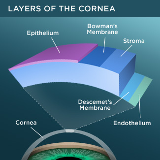
I am currently using radially acquired anterior segment OCT images of the cornea.

Goals

My goals are to segment the corneal layers

Methods I, IP

Graph-based segmentation - integrating soft surface-set feasibility constraints, possibly using arc-based graph search approach to support preferred direction, .perhaps use a flattened image to simplify the curving surface issue.

Methods II, AI

Use region-based costs - trained using random forest ... make sure it is different from earlier similar approaches.

Validation

Independent standard on 20 (30 is better) volumes - traced by VR (perhaps not all slices traced, but at least 300 slices traced ... and approved (hopefully) by MKG, make sure to fully separate training and testing - perhaps use leave one out.


Name: (John) David Stobaugh

Image Data

Encoding logs from HEVC(h265) Test Model.

Goals

Develop an improved Coding Unit "Type" decision for HEVC

Methods I, IP

Develop a meaningful automated metric of human-perceived image quality based on more information from a CU and from the pre-processing of a frame


Methods II, AI

Experiment with different classifiers to try to find a way to have an approach train itself for a video segment to better suit the underlying image data then retrain on scene transition


Validation

Given a finite number of bits, increase frame PSNR and improve visual clarity for the user. Assess in at least 15 image sequences.

Validation has to have 2 stages:

1) validating the automated metric of perceived image quality to show that it indeed reflects image quality.
2) Assume that the metric is validated, show that using your new approach (Method II) gives a better/comparable/worse results than current satndard encoding (run through standard HEVC (HM12.1) encoder)


Name: Ethan Ulrich

Image Data

3D PET/CT datasets (FDG and/or FLT) of head and neck cancer patients.  Pre- and post-treatment scans.

Goals

Automatically identify centers of uptake regions in the post-treatment PET scan.

Methods I, IP

Unsupervised registration of pre- and post-treatment scans.  Graph-based approach to search for best center.

Methods II, AI

Use a priori knowledge of the regions of interest from the pre-treatment scan to help with the initial placement of the corresponding centers in the post-treatment scan.

Validation

Using at least 30 datasets, compare with centers placed by expert given the same a priori knowledge.

Name: Markus VanTol

Image Data

Human head/neck PET scans with target locations for the lymph nodes

Goals

Segment multiple lymph nodes in a chain or cluster without a bias toward any of them.

Starting with 2 nodes, maybe go with 3 or more,

Methods I, IP

Watershed algorithm, thresholding, various other graph-based methods to find the gaps within and edges of the clusters, mesh voxelization.

The issue with my current method is that the segmentation is slightly different depending on the order of segmentation, so the idea of this would be to come up with a method that removes that bit of variation by segmenting the parts simultaneously.

Methods II, AI

Optimal Surface Finding with multiple graph centers; uses maxflow to efficiently determine the boundaries.

Validation

Method results will be compared in at least 20 hot spots to physician's segmentations of the same clusters. Quantitative comparisons such as the Dice coefficient and Jaccard index will be the main method of validation.

Name: Jin Wang

Image Data

Video sequences of stationary or moving cameras containing moving objects as foreground

Goals

Segment the foreground from the background of video sequences

Methods I, IP

Frame alignment and preprocessing - to be fine-tuned.

Methods II, AI

Train a dictionary and construct a convex optimization model to decompose the frames, as the foreground of moving objects can usually be sparsely represented.

Validation

Use 20 standard test video sequences with at least 50 frames each to validate the scheme both subjectively and objectively. Make sure the training and testing are completely separated.


Last Modified: 2014

[Go Back]