This page provides a short overview about our current work on the geometrically correct reconstruction of IVUS image sets by fusion with biplane angiography.
Also visit the Grant Web Page at the UI College of Engineering Imaging Group for the NIH-R01 grant HL63373 (Java required, last updated 1999)
Describes the purpose, methods, and early results of our approach as of 1998 - please refer to the posters and slide shows below for additional and more current information (HTML, 340 KB)
Medical Image Computing and Computer-Assisted Intervention 1998, presenting a comprehensive overview about the fusion system and its validation (PDF, 1867 KB)
SPIE's Medical Imaging Conference 1999, where a detailed description of our new approach for establishing the absolute orientation of the IVUS frames was presented - this poster received a Honorable Mention Poster Award of the Image Processing Conference. (PDF, 819 KB)
Computational Hemodynamics and Virtual Angioscopy, a collection of some screendumps showing the CFD analysis of a stenosed RCA in-vivo and the new interactive virtual angioscopy library implemented in VRML and JavaScript [based on data derived in cooperation with the University Hospital Essen, Germany, and the Brigham and Women's Hospital as well as Northeastern University in Boston, Massachusetts] (PDF, 1580 KB)
SPIE's Medical Imaging Conference 2003, presenting an application of the fusion system to assess the results of intravascular brachytherapy in coronary arteries based on a distance model, measured in four layers of the vessel wall - this paper was also the basis for an IEEE Transactions on Biomedical Engineering paper, see Publications section. (PDF, 4243 KB)
SPIE's Medical Imaging Conference 2005, providing results on the analysis of relationships between plaque distribution and vessel geometry (curvature) as well as hemodynamics (wall shear stress); results suggest that they correlate in the onset of atherosclerosis disease, but that hemodynamics is distorted once compensatory enlargement (positive remodeling) is exhausted and lumen narrowing occurs. (PDF, 2227 KB)
Note: For best impression, view PDF files in full-screen mode ([CTRL-L] in Acrobat-Reader)
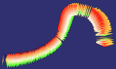
Scene which shows the angiographic setup as well as the spatially reconstructed path of the catheter and the IVUS images at their correct locations and in their correct orientations:
Scene which shows the segmentation results:
Red Inner border of the wall, and
Green Outer border of the wall.
A virtual angioscopy animation based on
a set of automatically generated viewpoints (without JavaScript support
for interaction) allows to "fly through" the vessel
-
for best impression, follow the viewpoints backwards.
For general information about the Virtual Reality Modeling Language, refer to the VRML-Consortium Home Page.
The scenes were generated from in-vitro data, which was obtained using an undiseased cadaveric pig heart.
There is further information given by the list of publications and by further images.
Please use at least an HTML 3.0 viewer such as Netscape 4.x for tables and figures, and SGI CosmoPlayer 2.0 or comparable for VRML 2.0 scenes!